CONFLUENCE
the confluence challenge
Confluence indicates the percentage of the surface to which cells adhere to (e.g. in culture flask or a well on a multi-well plate), as compared to the free space, not covered by cells. For example, 75 % confluence means that 75 % of the surface is covered by cells, while 25 % is still free.
Confluence is one of the crucial parameters for monitoring cells growing in monolayer in vitro. It helps to control the speed of the culture expansion and optimal time for the next passage. In order to ensure the good health of the cells and reproducibility of the experiments the confluence should not be neither too low nor too high. The optimal confluence window and the sensitivity of cells to its deviation, depend on specific cell type.
A smart and accurate solution
Confluence measurement is often done using a naked eye. This is however a rough estimation.
OUR SOLUTION
We have developed a tool for automatic and accurate assessment of exact confluence based on the phase microscopy image. This tool is successfully used for routine checks of neuronal cell cultures at Final Spark lab.
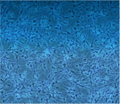
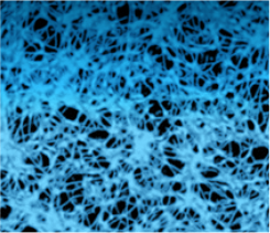
Our software is based on U-Net architecture, applicable for image segmentation. During the analysis, a UNET map is created, as on the image below. According to this map, computer ‘recognises’ which pixels on the image represent cells. Based on this, a percentage of pixels represented by the cells, as compared to the whole surface, is calculated.
The result is a process that is:
- much more accurate
- extremely time saving

image of cells

unet map

accurate result
Confluence
Let’s talk about your own challenge – and let us propose a solution that works for you.
try this demo
HOW CAN WE HELP YOU?
Give us a few details and we’ll contact you.
Or call to find out more
